Receptor
A receptor is a molecule usually found on the surface of a cell that receives chemical signals from outside the cell. When such external substances bind to a receptor, they direct the cell to do something, such as divide, die, or allow specific substances to enter or exit the cell. A molecule that binds to a receptor is called a ligand and can be a peptide or another small molecule such as a neurotransmitter, hormone, pharmaceutical drug, or toxin.
Structure
Receptors are proteins embedded in either the cell's plasma membrane, in the cytoplasm, or in the cell's nucleus to which specific signaling molecules may attach.
- The primary structure of a protein is the order of amino acids that make up that polypeptide chain.
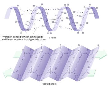
- The secondary structure of a protein is the folding of the amino acids of the polypeptide into different shapes. These are often α-helices or β-pleated sheets. They are held together by hydrogen bonds, which increase the stability of the protein.
- The tertiary structure of a protein is the final 3D structure of the protein. The tertiary structure is made up of four different bonds or interactions; disulphide bonds, ionic bonds, hydrogen bonds and hydrophilic/hydrophobic interactions ("love"/"hate" of water).
- The quaternary structure of a protein is when a protein is made up of several polypeptide chains and sometimes an inorganic molecule, known as a prosthetic group.
Binding
Numerous receptor types are found in a typical cell. Each type is linked to a specific biochemical pathway and binds to only certain ligand shapes. When a ligand binds to its corresponding receptor, it activates or inhibits the receptor's associated biochemical pathway.
Lock and key model
It has been suggested that both the protein and the ligand possess specific complementary geometric shapes that fit exactly into one another. This is often referred to as "the lock and key" model.
Induced fit model
The induced fit model suggests that the active site of the receptor will bind to the ligand and then alter its 3D shape to perfectly contain the ligand while reacting. Once the reaction is complete, the 3D shape will revert to normal and the metabolites of the ligand are released. This is generally the more accepted theory of binding.
Activation
Ligand binding changes the three-dimensional shape of the receptor molecule. This alters the shape at a different part of the protein, changing the interaction of the receptor molecule with associated biochemicals, leading in turn to a cellular response mediated by the associated biochemical pathway. However, some ligands called antagonists merely block receptors from binding to other ligands without inducing any response themselves. The action of ligands can be measured via two methods; the ability of the drug to combine with the receptor to create drug-receptor complex (affinity) and the ability of the drug-receptor complex to initiate a response (efficacy).
Inhibition of activation
The blockade of a specific receptor type or subtype is facilitated by chemicals known as antagonists. There are several different types of antagonists. A competitive antagonist is a receptor antagonist that binds to a receptor but does not activate the receptor. The antagonist will compete with available agonist for receptor binding sites on the same receptor. A sufficient antagonist will displace the agonist from the binding sites, resulting in a lower frequency of receptor activation. Another way antagonists work is by blocking the channels, which were opened by the ligand activation because of their chemical shape. These are called uncompetitive antagonists because no matter how much ligand is present, the channel is still blocked and the ligand activation of the receptor does nothing (DXM is a common example).
Ion channels
Some receptors, such as the GABAA receptor, are ion channels. Ion channels are proteins that are situated on the cell membrane and allow the transportation of ions across it. For example, the GABAA receptor is a chlorine ion channel, meaning it transports charged chlorine (Cl-) ions across the cell membrane of the neuron.
Generally, ion channels are divided into two categories, voltage-gated and ligand-gated. Voltage-gated ion channels are activated by electrical charges. This means that when the electrical charge near the channel changes, the channel will let ions through. On the other hand, the other type of ion channel, ligand-gated ion channels, are activated by a chemical (often a neurotransmitter).
The GABAA receptor is ligand gated. Whenever a gamma-aminobutryric acid molecule binds to the receptor, the receptor will allow chlorine ions to pass through. Ion channels are extremely common throughout not only the brain, but also the body. For example, the NMDA receptor is a potassium ion channel and the drug verapamil blocks calcium channels to treat hypertension and various heart diseases.
See also
External links
References
This article does not cite enough references. You can help by adding some. |
